DIABETIC RETINOPATHY
What is the diabetic retinopathy?
The diabetic patients have increased sugar levels in their vessels of the whole body, including the eye. High glucose results in damage of the wall of the vessels and that is the initial factor of the whole complications: bleedings, lipid exsudates, ischemic areas, new leaking vessels, swelling of the macula etc. The severity of the disease may rank from a mild form to a very severe one and so are the symptoms, mild, moderate or even blindness.
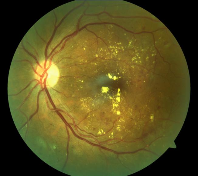
Non proliferative diabetic retinopathy with bleedings and lipid deposits
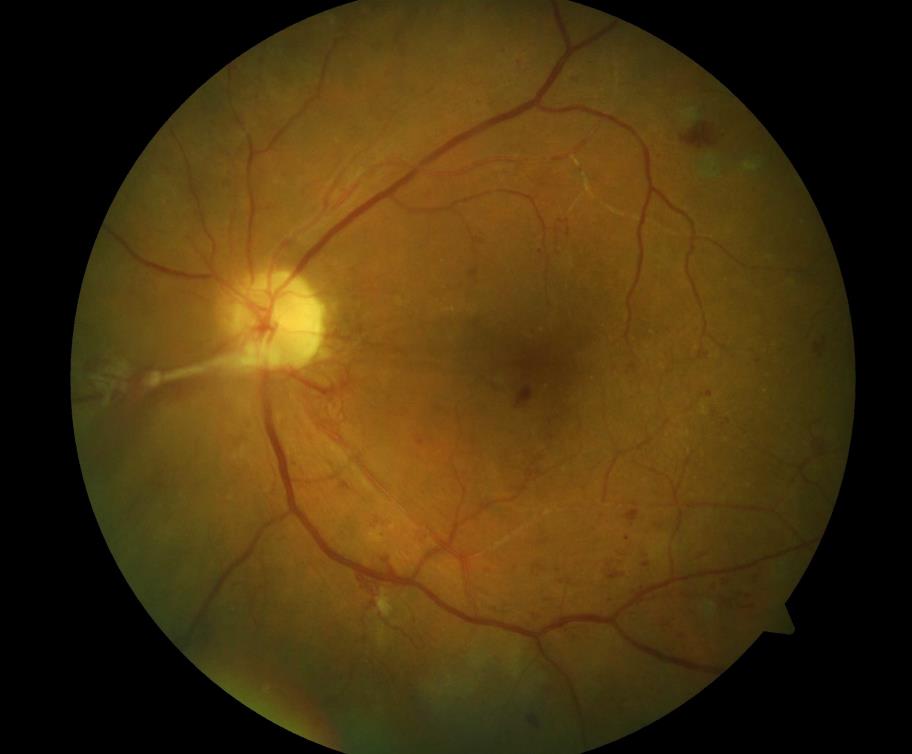
Proliferative diabetic retinopathy with new vessels on the optic disc

The left photo is the normal one and on the right is how someone with diabetic retinopathy sees
The diagnosis of the diabetic retinopathy
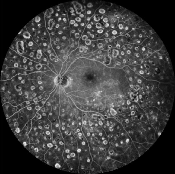
The most important is the examination of the retina of all diabetic patients once a year. The retina should be examinated thoroughly, after dilatation of the pupil with special eye drops. This control examination should be more often, if the diabetic patient has symptoms or if his sugar levels are not well regulated. If somebody has already a diabetic retinopathy, two are the most crucial examinations: the OCT of the macula ( in order to see if there is macular oedema) and the fluorescence angiography ( in order to see if there are ischemic areas of the retina).
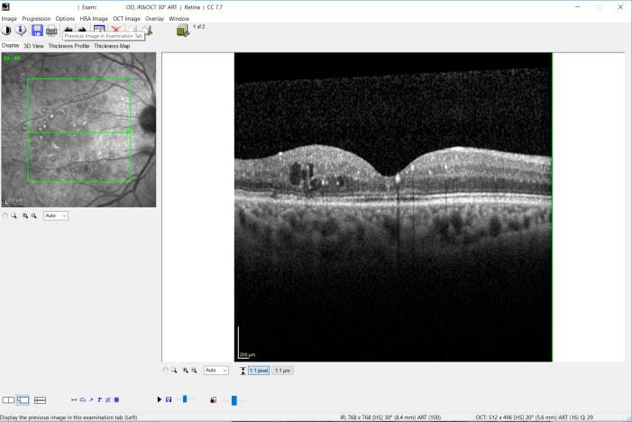
the OCT of a macula with diabetic macular oedema
The treatment of the diabetic retinopathy
The treatment of the diabetic retinopathy is different, according to the findings of the OCT and the fluorescence angiography. If there is macular oedema, the treatment of choice are intravitreal injections with special medications. If there are ischemic areas, we use the argon laser to coagulate them. An optimal regulation of the blood sugar levels is always essential, both as a prevention measurement and as a treatment choice.
A photo of a retina after argon photocoagulation